Back Of Neck Anatomy Glands - The Lymphatics Of The Head Face And Neck Human Anatomy : Swollen neck glands that persist for a long time are known as chronic swollen glands in neck.
Back Of Neck Anatomy Glands - The Lymphatics Of The Head Face And Neck Human Anatomy : Swollen neck glands that persist for a long time are known as chronic swollen glands in neck.. Related posts of anatomy of neck muscles. Such a division has an anatomical substrate, because the deep fascia of the neck sends in frontal 8. Anatomy, head and neck, palatoglossus muscle (glossopalatinus. Learn about glands salivary neck anatomy with free interactive flashcards. Sometimes a pyramidal lobe is also present, extending upward anterior to the thyroid cartilage.
Anatomy of the human body. the strap muscles are retracted to access the. The head rests on the top part of the vertebral column, with the skull joining at c1. And then you've got the prevertebral fascia, which actually runs all the way back to enclose the vertebral column and the muscles associated with it. Sometimes a pyramidal lobe is also present, extending upward anterior to the thyroid cartilage.
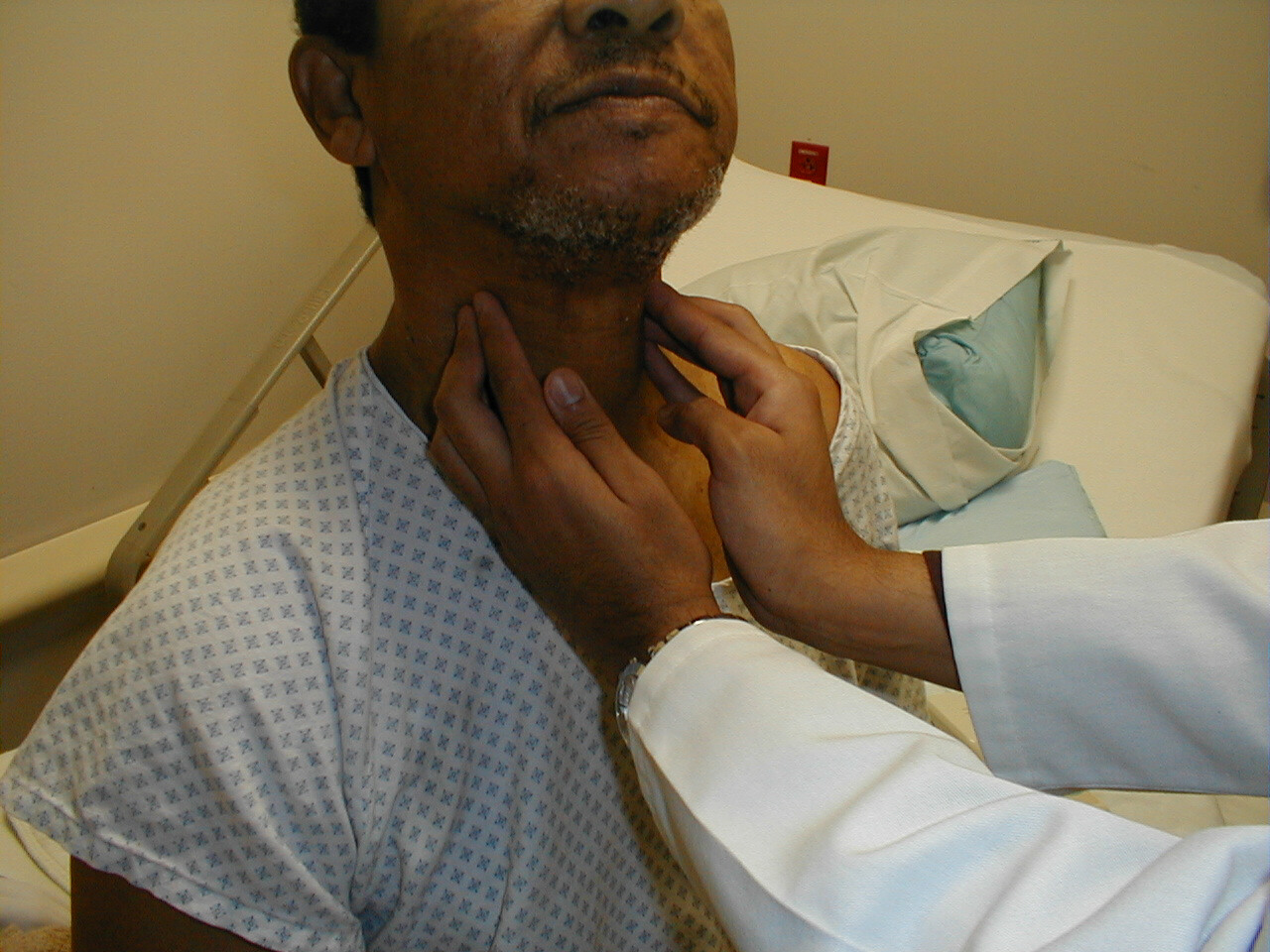
Uc San Diego S Practical Guide To Clinical Medicine from meded.ucsd.edu In some cases, inflammation of neck glands may occur due to hodgkin's. It runs down the back part of the neck, and opens into the external jugular vein just below the middle of its course. Anatomy of the human body. Despite being a relatively small region, it contains a range of important anatomical features. There are lymph nodes on the back of the neck which may become inflamed with infections both viral and bacterial. Normally, the thyroglossal duct then involutes, but when the duct persists, a thyroglossal duct cyst can develop anywhere along this tract (figure). Jugularis 560) begins in the substance and on the surface of the thyroid gland, by tributaries corresponding with the branches of the superior thyroid artery, and. Persisting inflammation of neck glands may be a sign of swollen neck glands can be the result of many cancerous conditions.
Jugularis 560) begins in the substance and on the surface of the thyroid gland, by tributaries corresponding with the branches of the superior thyroid artery, and.
A collection of anatomy notes covering the key anatomy concepts that medical students need to learn. Sometimes a pyramidal lobe is also present, extending upward anterior to the thyroid cartilage. 803 x 1024 jpeg 192 кб. The superior fibres draw the tip back rathee m, jain p. The endocrine system includes all of the glands of the body and the hormones produced by those glands. We've also got the parathyroid glands behind the thyroid. Anatomy of the human body. Lablink anterior neck & thorax. The anterior jugular vein (v. Normally, the thyroglossal duct then involutes, but when the duct persists, a thyroglossal duct cyst can develop anywhere along this tract (figure). Jugularis 560) begins in the substance and on the surface of the thyroid gland, by tributaries corresponding with the branches of the superior thyroid artery, and. Major glands are the primary glands providing the oral cavity and its structure moistening, lubrication, and protection. Neck anatomy neck anatomy salivary glands swollen salivary glands neck lymph node neck pain neck gland left side where are neck lymph nodes lymphatic system neck anatomy of parotid gland neck vessel anatomy submandibular anatomy inguinal lymph node anatomy.
There are lymph nodes on the back of the neck which may become inflamed with infections both viral and bacterial. Cervical fascia and interfascial spaces in the neck. Despite being a relatively small region, it contains a range of important anatomical features. The 5 anatomical spaces of the infrahyoid neck. Youtube makes it easy to share.
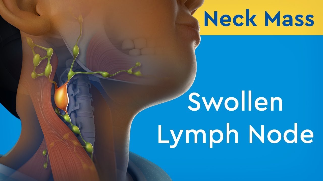
Causes For Swollen Lymph Nodes In The Body Thehealthsite Com from st1.thehealthsite.com Anatomy of the human body. Anatomy of neck spaces and levels of cervical lymph nodes by dr. 803 x 1024 jpeg 192 кб. Lablink anterior neck & thorax. The anatomy of the head and neck is complex because so many different functional structures are located close to each other. Such a division has an anatomical substrate, because the deep fascia of the neck sends in frontal 8. Level ii upper internal jugular nodes, posterior to the back of the submandibular salivary gland, anterior to the back of the sternocleidomastoid muscle. This article describes the anatomy of the head and neck of the human body, including the brain, bones, muscles, blood vessels, nerves, glands, nose, mouth, teeth, tongue, and throat.
Anatomy, head and neck, palatoglossus muscle (glossopalatinus.
Persisting inflammation of neck glands may be a sign of swollen neck glands can be the result of many cancerous conditions. The embryonic thyroid gland or thyroid anlage travels through the duct to reach its final normal position. The anterior jugular vein (v. Thyroid gland, parathyroid glands, larynx, trachea, esophagus, submandibular gland, caudal part of the parotid gland. Anatomy of neck spaces and levels of cervical lymph nodes by dr. Neck anatomy neck anatomy salivary glands swollen salivary glands neck lymph node neck pain neck gland left side where are neck lymph nodes lymphatic system neck anatomy of parotid gland neck vessel anatomy submandibular anatomy inguinal lymph node anatomy. Learn about glands salivary neck anatomy with free interactive flashcards. The neck is a complex anatomic region between the head and the body. the strap muscles are retracted to access the. Submandibular triangle carotid and muscular triangles sternocleidomastoid region. Swollen neck glands that persist for a long time are known as chronic swollen glands in neck. I thought i'd use this channel to share some anatomy thoughts and include some of the other stuff too. The head rests on the top part of the vertebral column, with the skull joining at c1.
Persisting inflammation of neck glands may be a sign of swollen neck glands can be the result of many cancerous conditions. The neck is a complex anatomic region between the head and the body. I thought i'd use this channel to share some anatomy thoughts and include some of the other stuff too. The thyroid gland consists of two lateral lobes joined by an isthmus. 803 x 1024 jpeg 192 кб.
Neck Mass Swollen Lymph Node Youtube from i.ytimg.com Learn about glands salivary neck anatomy with free interactive flashcards. The neck is the area between the skull base and the clavicles. Superior and inferior thyroid, common carotid, external carotid, internal carotid artery (and sinus), facial, submental, lingual arteries. Head and neck anatomy is important when considering pathology affecting the same area. The thyroid gland consists of two lateral lobes joined by an isthmus. Sometimes a pyramidal lobe is also present, extending upward anterior to the thyroid cartilage. Level ii upper internal jugular nodes, posterior to the back of the submandibular salivary gland, anterior to the back of the sternocleidomastoid muscle. Normally, the thyroglossal duct then involutes, but when the duct persists, a thyroglossal duct cyst can develop anywhere along this tract (figure).
The thyroid gland consists of two lateral lobes joined by an isthmus.
Cervical fascia and interfascial spaces in the neck. The 5 anatomical spaces of the infrahyoid neck. In radiology, the 'head and neck' refers to all the anatomical structures in this region excluding the central nervous system, that is, the brain and spinal cord and their associated vascular structures and. Despite being a relatively small region, it contains a range of important anatomical features. The embryonic thyroid gland or thyroid anlage travels through the duct to reach its final normal position. 3.6) and 120° in the female (fig. A collection of anatomy notes covering the key anatomy concepts that medical students need to learn. Normally, the thyroglossal duct then involutes, but when the duct persists, a thyroglossal duct cyst can develop anywhere along this tract (figure). Head and neck anatomy is important when considering pathology affecting the same area. The vocal cords are attached to the back of this prominence, and muscles attached to the oblique line, on the outer surface of the cartilage, to the. Anatomy and function neck, regions of the lower face, cervical spine, head joints,.during muscle traction, the cheeks are pulled together, which makes food move back and forth between the.the parotid gland, which is one of the 3 major salivary glands. 803 x 1024 jpeg 192 кб. Trachea and thyroid gland and also form the anterior boundaries of the neck levels.
Thyroid gland, parathyroid glands, larynx, trachea, esophagus, submandibular gland, caudal part of the parotid gland back of neck anatomy. It runs down the back part of the neck, and opens into the external jugular vein just below the middle of its course.


0 Komentar